In July we gathered at the Royal Free Hospital for a collection of talks on COPD, asthma, NTMs and a focus on pleural disease.

Spotted this Patrick Caulfield on the walls of the Royal Free?
Academic COPD update and the new London α1 antitrypsin deficiency
Dr John Hurst began the day with a review of COPD, encouraging us to question the current methods of defining and diagnosing COPD, and to be more precise in our approach.
He asked the audience what is the current definition of COPD?
- smoking + genetic susceptibility = COPD
- Emphysema = pathological diagnosis of destruction of air spaces
- Chronic bronchitis = clinical definition of chronic mucus production for at least 3 months in two successive years
- Physiological diagnosis post bronchodilator FEV1/FVC <0.7
He went on to explore what a COPD phenotype is: a gene-environment interaction leading to “a single or combination of attributes that describe differences between individuals with COPD as they relate to meaningful outcomes”. Bronchodilator reversibility is not a phenotype as it is not stable (Calverley, P. M. A., et al. “Bronchodilator reversibility testing in chronic obstructive pulmonary disease.” Thorax 58.8 (2003): 659-664).
Severity is based on the GOLD criteria, combining symptom burden, degree of airflow limitation and exacerbation frequency. Treatment is determined by stage (ICS, LABA and LAMA). We noted that 2011 GOLD was pharma-sponsored, under the auspices of the WHO, which may or my not influence guidance. GOLD utilises: MRC breathlessness scale; CAT (available in many languages); RISK GOLD stage (spirometry); and exacerbation frequency
What is an exacerbation?
- viruses – ie Sebastian Johnston experimental model of rhinovirus-induced COPD exacerbation (Mallia, Patrick, et al. “Experimental rhinovirus infection as a human model of chronic obstructive pulmonary disease exacerbation.” American journal of respiratory and critical care medicine 183.6 (2011): 734-742.)
- bacteria – change in the microbiome post-viral infection in COPD ( Dickson, Robert P., et al. “The lung microbiome and viral-induced exacerbations of chronic obstructive pulmonary disease: new observations, novel approaches.” American journal of respiratory and critical care medicine188.10 (2013): 1185-1186.)
- change in strain H influenzae, M catarrhalis, or Strep pneumoniae (Sethi, Sanjay, et al. “New strains of bacteria and exacerbations of chronic obstructive pulmonary disease.” New England Journal of Medicine 347.7 (2002): 465-471.)
- not related to bacterial load (Monso, E., et al. “Bacterial infection in chronic obstructive pulmonary disease. A study of stable and exacerbated outpatients using the protected specimen brush.” American journal of respiratory and critical care medicine 152.4 (1995): 1316-1320.)
A review on the lung microbiome in COPD is available in AJRCCM: Gosselink, J. V., et al. “The lung microbiome in COPD.” Am J Respir Crit Care Med 183 (2011): A1017.
Dr Hurst challenged our understanding of COPD and suggested future areas of research. In particular he asked what determines susceptibility to an exacerbation. He reminded us that the best predictor of future exacerbation is previous exacerbation number.
He also highlighted the London α1-antitrypsin deficiency service which provides joined-up care (hepatology and respiratory input), expert advice, training and research. Referrals are welcomed from any hospital. Patients who are found to be homozygous, or those with difficult phenotyping should be referred. If you’re interested in getting involved in research, get in touch.
Key messages regarding COPD were:
- diagnose COPD only with spirometry
- there is currently lots of (too much?) interest in phenotyping
- exacerbations are caused by viruses and alteration in bacterial flora – future treatment/prevention strategies?
- manage cardiovascular risk and you might save a life ( raised troponin and BNP x9 risk mortality at 30 and 90 days so refer for appropriate tests eg MPS in order to make sure patients receive interventions for reversible ischaemia)
- get the basics right!
Slides are available here: HURST_COPD_Handout
NTM: what should we do about it?
Dr Marc Lipman began with an honest summary of the evidence-base and guidelines related to Non Tuberculous Mycobacteria: it’s difficult! Most guidelines are based on expert opinion, case series and a small number of RCTs.
NTMs are environmental mycobacteria that do not cause TB or leprosy. The clinical syndromes include:
- Pulmonary disease: MAC, kansasii
- Lymphadenitis: seen mainly in children<5yrs ie scrofulaceum, malmoese, haemophilum (highly curable)
- Disseminated: MAC
- Soft tissue: abscessus, chelonae, fortuitum, ulcerans, xenopi, marinum
One of the reasons that it is difficult to provide clear guidance on NTMs is that there is a lack of data on prevalence, particularly in certain parts of the world eg Africa. This paper demonstrated that the species distribution among NTM isolates from pulmonary specimens differed by continent, and by country within these continents.
The reporting of NTM is not mandatory (as it is for MTB) but laboratories voluntarily supply data to Public Health England. New data from PHE (as yet unpublished) provides a picture of England and Wales 2007-12. Isolates were 33% M. avium-intracellulare, 11% M. chelonae, 14% M. kansasii. There was in increased prevalence with age. In this data M. abscessus was associated with a CF population, and therefore affected more young people, chelonae was associated with line infections and xenopi was found in older patients (men more than women but this may represent confounding from the gender difference in COPD prevalence).
“Lady Windermere syndrome” is the name for pulmonary infection with MAC, named after a character in the Oscar Wilde play. The lacy structure of the infection may also have influenced the name.
So, once we identify an NTM, what should we do?
ATS/IDSA guidelines suggest indications for treatment of pulmonary infection are:
- respiratory symptoms and radiographic abnormalities (nodules, cavities, multifocal bronchiectasis)
- PLUS exclusion of other diagnoses
- PLUS >2 positive cultures OR 1 positive bronchial washing culture OR 1 positive sputum culture and histological confirmation of pulmonary involvement
They also highlight the fact that making the diagnosis of NTM lung disease does not per se necessitate the institution of therapy, a decision which should be based on the risks and benefits for the individual patient. These are pragmatic guidelines based on a small amount of evidence.
The ATS and BTS guidelines differ in terms of treatment regimes, as BTS does not include a macrolide for MAC. In fact there is poor evidence for all drugs and combinations. A recent review collates the evidence:
On the basis of this, they recommend RE-macrolide +/- AS (Rifampicin, Ethambutol and a Macrolide with or without either Amikacin or Streptomycin) until culture negative for >12 months. The addition of streptomycin is debated as compliance is difficult for this duration of treatment and, although a trial showed greater efficacy for this regime follow-up was not as long as for other trials.
The January edition of Annals of the ATS has a special feature on NTMs, including a call from Dr Lipman and others for a pragmatic, patient-centred approach to care. They advocate for new research studies, including patient-reported experience measures and patient-reported outcome measures which they hope will enable patients and their health-care providers to make clinical management decisions that derive from a realistic view of what they can hope to achieve from treatment.
Slides are available here: Resp North Thames Trainees NTM what to do Marc Lipman handout 7 July 2014
Pleural fluid myths and elusive effusions
Dr James Goldring was straight after coffee, with a perfectly pitched exploration of pleural effusions. Who knew there was so much more to it than Light’s criteria….?
James began by challenging some of our assumptions, and highlighted that 9% of transudates were turbid, and only 11% of malignant effusions were blood stained in one series (overall 47% of bloody effusions were malignant). Don’t always believe your eyes.
He reminded us to consider chylothorax as the diagnosis can be missed without a high index of clinical suspicion. Only a minority are ‘milky’ as is classically described. If suspected the fluid should be analysed for triglycerides, but 14% had triglycerides <1.2mmol/L (hence traditional triglyceride cutoff values used in excluding the presence of chylothorax may miss the diagnosis in fasting patients, particularly in the postoperative state). Other useful tests are chylomicrons and lipoprotein electrophoresis.
The optimal volume of fluid for cytology is often debated. Although guidelines suggest that there is no addition value to sending >60ml, it is possible for labs to create a cell block from larger volumes (ie 150ml) which then increase the pickup rate for malignancy on pleural fluid samples. If in doubt, call your lab. Another great tip was that if there is a delay in the ability to process a cytology sample (eg over a bank holiday) it will be fine in a fridge for up to 14 days.
- Swiderek, Jennifer, et al. “Prospective study to determine the volume of pleural fluid required to diagnose malignancy.” CHEST Journal 137.1 (2010): 68-73.
- Abouzgheib, Wissam, et al. “A prospective study of the volume of pleural fluid required for accurate diagnosis of malignant pleural effusion.” CHEST Journal135.4 (2009): 999-1001.
Light’s criteria was revisited, and we were reminded it is 98% sensitive 74% specific for identifying an exudate. Reassuringly, the paired serum sample can be up to 5 days after the pleural fluid sample – so don’t accept any excuses from referring teams!
Misclassification of transudates by Light’s criteria occurs in CCF (30%), and hepatic hydrothorax (18%), influenced by diuretic use.
In the case of CCF we were advised to use the albumin gradient: serum albumin-pleural albumin >12g/L which correctly identified 83% of ‘false exudates’ (see Bielsa, Silvia, et al. “Solving the Light’s criteria misclassification rate of cardiac and hepatic transudates.” Respirology 17.4 (2012): 721-726.) We then considered the use of NT-proBNP. Serum and pleural levels are highly correlated so it is probably wise to just to a serum level. The only studies that have looked at pleural levels have used very high levels.
We considered possible explanations for discordant or unusual results:
- high protein, low LDH: bloody tap, chylothorax, yellow nail syndrome
- high LDH, low protein: malignancy, parapneumonic effusion, PCP
- very high LDH >1000: infection >> malignancy > RA
- very high protein >70g/L: multiple myeloma, waldenstroms macroglobulinaemia
Despite the usual investigations as per BTS guidelines, between 5-20% of unilateral pleural effusions remain undiagnosed. This may even be the case after thoracoscopic biopsy (Venekamp, L. N., B. Velkeniers, and M. Noppen. “Does ‘Idiopathic Pleuritis’ exist? Natural history of non-specific pleuritis diagnosed after thoracoscopy.”Respiration 72.1 (2005): 74-78.). Possible uncommon causes include urinothorax and drug induced effusion (see pneumotox). In addition PE may be a more common cause than we think – perhaps the 4th most common cause? In fact it is usually no more than a blunted costophrenic angle, and does not usually persist beyond 3/52. Reassuringly most undiagnosed unilateral effusions are benign ((91% in the above study)) and resolve (81% in the above study). Therefore unless there are other concerning features a ‘watch and wait’ policy is safe.
NB. since this training day a paper in Thorax has sought to predict survival in malignant pleural effusion using the LENT score, claiming greater accuracy than ECOG status alone (Clive, Amelia O., et al. “Predicting survival in malignant pleural effusion: development and validation of the LENT prognostic score.” Thorax (2014): thoraxjnl-2014). You can also read the rapid response to this article by NET trainee Dr James Murray et al, and the author’s response.
Slides are available here: Pleural fluid myths and elusive effusions2
Add on treatments in asthma
Next Dr Paul Dilworth, Nicola Marks and Lauren Geddes discussed the Royal Free’s experience of omalizumab, and the use of physiotherapy for dysfunctional breathing.
We were reminded that severe asthma is not only distressing and debilitating for patients, but also has significant direct and indirect costs for the UK. Omalizumab (or Xolair) is a recombinant DNA derived humanised IgG1k monoclonal antibody which binds circulating IgE, and is approved for those with severe asthma not adequately controlled with high dose corticosteroids. The INNOVATE trial (Humbert, M., et al. “Benefits of omalizumab as add‐on therapy in patients with severe persistent asthma who are inadequately controlled despite best available therapy (GINA 2002 step 4 treatment): INNOVATE.” Allergy 60.3 (2005): 309-316) showed a decrease in the rate of clinically significant asthma exacerbations, severe exacerbations, and hospital attendances. Results have since been replicated in ‘real-world’ studies. Anaphylaxis occurs in 0.09-0.2% of cases.
- Korn, S., et al. “Omalizumab in patients with severe persistent allergic asthma in a real-life setting in Germany.” Respiratory medicine 103.11 (2009): 1725-1731
- Barnes, Neil, and Amr Radwan. “The APEX study: Retrospective review of oral corticosteroid use in omalizumab-treated severe allergic asthma patients in UK clinical practice.” European Respiratory Journal 38.Suppl 55 (2011): p269
- Schumann, Christian, et al. “Omalizumab in patients with severe asthma: the XCLUSIVE study.” The clinical respiratory journal 6.4 (2012): 215-227
Criteria for use are:
- >6yrs old
- severe persistent confirmed allergic IgE-mediated (eg skin prick test proven) asthma with continuous/frequent use of oral corticosteroids (ie 4 courses in 12 months)
- optimisation of all other therapy
- IgE <1500
- impaired lung function
- other issues excluded ie ABPA, dysfunctional breathing
Outcomes from RCT and real world practice include decreased exacerbations, decreased A+E and hospital visits, decreased oral corticosteroid use, decreased symptom burden, decreased number of days off work, increased asthma-related quality of life and improved lung function. We were reminded of the scales used to measure symptoms over time, including the ACT and AQoLQ.
Trainees underestimated the impact of Omalizumab, and were encouraged to be less pessimistic!
Moving onto dysfunctional breathing Lauren Geddes, lead Respiratory physio described the complex interaction between organic, phychogenic and physiological factors. Symptoms are variable and multisystem:
- Resp: breathlessness, sighing, yawning, air hunger, unsatisfying deep breaths, dry cough
- CV: palpitations, chest pain, tachycardia
- Neuro: dizziness, paraesthesia, confusion, poor concentration, tetany (rare)
- GI: dyspepsia, bloating, reflux
- MSkel: cramps, spasms, neck pain, restless legs, postural abnormalities
- psych: anxiety, panic attacks, phobias, depression
These symptoms can create a vicious cycle of fear, deconditioning, isolation and worsening symptoms.
Assessment aids used by physios include:
- Nijmegin questionnaire to diagnose hyperventilation in asthma (score of more than 23 out of max 64 positive)
- Breath-hold test (on inhalation – 30sec is v good, 10-15sec suggests disordered breathing pattern)
- Hyperventilation provocation test (ETCO2 or ABG assessment)
- Clinical assessment including history, observation of breathing pattern, sleep quality, lifestyle, diet, triggers
The physical assessment includes observation of the speed, depth and co-ordination of breaths, diaphragm use, accessory muscle use, I:E ratio, breath-holding and breath-stacking, in addition to posture and lung volumes.
Some are described in this review: Vansteenkiste, Johan, F. Rochette, and Maurice Demedts. “Diagnostic tests of hyperventilation syndrome.” European Respiratory Journal 4.4 (1991): 393-399.
There is some evidence for the efficacy of breathing retraining:
- Thomas, Mike, et al. “Breathing exercises for asthma: a randomised controlled trial.” Thorax 64.1 (2009): 55-61. Led to improvements in asthma-specific health status and other patient-centred measures but not in asthma pathophysiology.
- Holloway, E., and F. S. Ram. “Breathing exercises for asthma.” Cochrane Database Syst Rev 1 (2004). Only 7 studies were included. Most studies were of small size. Two studies demonstrated significant reductions in rescue bronchodilator use and three studies showed reductions in acute exacerbations, although these were measured in different ways. Two single studies showed significant improvements in quality of life measures.
CBT has been used to address the psychological aspects, and other approaches including Buteyko breathing and alternative therapies are recommended by some, with little evidence.
Slides are available here: Asthma Symptom Scores; HYPERVENTILATION – A BREATH TOO FAR; Omalizumab
Medical Thoracoscopy
The afternoon was focused on pleural disease, and we began with a talk from Dr Alex West from across the river at St Thomas’ Hospital.
Dr West extolled the virtues of thoracoscopy, including the fact that you do not have to have surgeons on site. He reminded us that it was actually invented by a Swedish physician! If you are interested in becoming a thoracoscopist you can access training as a trainee at a number of centres, including Basildon Hospital.
We considered the great utility of ultrasound as part of a diagnostic and therapeutic service:
- identification of loculations
- visualisation of visceral pleural thickening
- pleural thickening amenable to targeted trucut biopsy
Dr West described what could be achieved with thoracoscopy when you were just starting to use the technique (more advanced skills required more experience):
- complete aspiration of effusion
- parietal pleura biopsy under direct vision
- breakdown of simple loculations
- talc poudrage and drain placement
Post talc poudrage the patient should be on suction for 48hours. Remember to omit NSAIDs and steroids. Pleurodesis can then be confirmed with ultrasound (lack of sliding lung sign). Port site radiotherapy is necessary if mesothelioma is confirmed to prevent seeding. This is irrespective of performance status.
Trapped lung can be difficult to diagnose in advance but is often evident at the time of the procedure. Insertion of a tunnelled indwelling pleural catheter should then be considered.
Thoracoscopy has advantages over VATS:
- no need for GA (so those with higher ASA scores are not excluded)
- day case or short stay (vs 4-5 days for VATS)
- well tolerated in the elderly
- cheaper as no need for anaesthetist or theatre (done in bronchoscopy suite)
Since ‘medical’ thoracoscopy is so great, who should be referred to the surgeons for VATS?
- ‘patient choice’ ie if anxious about awake procedure, likely to be poorly compliant (remember they will have to be fit for GA)
- malignant trapped lung in order to perform decortication
- complex empyema (although the debate rages about optimal use of tPa and DNAse)
- recurrent pneumothorax
- visceral pleural biopsy or wedge
Slides are available here: Medical Thoracoscopy – SpRs.ppt1
The remainder of the afternoon involved practical sessions: handling the kit for thoracoscopy; inserting indwelling pleural catheters on models; and enhancing our suturing skills under the guidance of the RFH plastics team. If you missed out on these great hands-on sessions have a look at these videos to get an idea of the procedures:
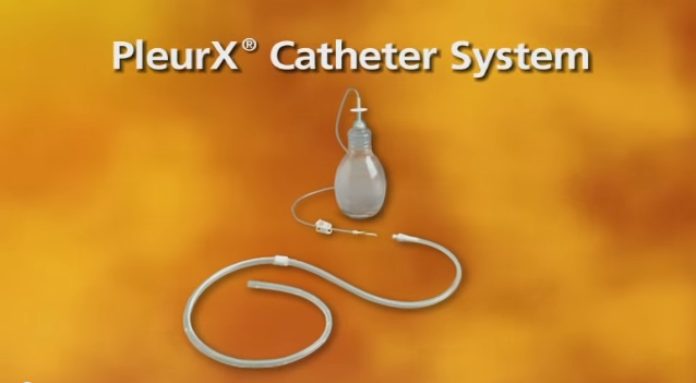
Other indwelling catheter systems are available…
- PleurX catheter insertion (try to ignore how American the video is!)
- Thoracoscopy
- Suturing chest drains

Discussion
No comments yet.