In April we convened at the North Middlesex Hospital for a diverse day of all things from immunohistochemistry to writing a business case via IGRA testing and interventional bronchoscopy.
Interesting facts about the North Middlesex Hospital:
- The North Middlesex Hospital began life as a workhouse in 1842, built by the Edmonton Board of Guardians.
- Every day, on average, the North Mid sees 500 patients in A&E; 15 babies are born in its’ maternity unit; about 450 inpatients are cared for on its’ wards; about 50 patients have major or minor surgery in one of its’ 10 operating theatres; and about 800 people attend the outpatients clinics
- The video for the Lostprophets song Shinobi vs. Dragon Ninja was filmed in a car park at Edmonton Green, near the North Middlesex Hospital.
- Famous people from the Edmonton area include Robert Cecil (first Earl of Salisbury), Bruce Forsyth (entertainer), and Florence Green (last surviving First World War veteran, who lived to 110).

Rarities of Respiratory Radiology
We were welcomed by Dr Bryan Sheinman, and then began with a session from Dr Sashin Kaneria on tips for interpreting respiratory imaging studies, particularly in identifying rare diseases. Our first case had a CT showing upper lobe predominant, patchy, subpleural reticular infiltrates. We considered a differential including TB, sarcoidosis and hypersensitivity pneumonitis. In fact an SpR was able to identify this as Idiopathic Pleuroparenchymal Fibroelastosis – a new ILD (example case report). 100 cases had been identified up to 2013. The median age is 57. Pneumothorax is a common complication. Recurrent infections occur in 50%. Clubbing is rare. Crackles are less common than in IPF (<50%).
Next we revised our knowledge of the CT Halo sign, also known as ‘Atoll’ sign. See Radiopaedia for examples.
- infection: invasive aspergillosis, coccidiomycosis, TB, pneumocystis pneumonia,
- neoplasm: adenocarcinoma in situ (previously known as BAC)
- other: post-radiotherapy/radiofrequency ablation, Wegeners, sarcoidosis
We were encouraged not to dismiss ground glass nodules, as some may be early lung cancers (particularly adenocarcinoma in situ, previously known as BAC). New Fleischner guidelines provide guidelines on how often to repeat CTs in part-solid part-ground glass and entirely ground glass nodules (handy app recommended in resources).
We were shown a CT showing ‘crazy-paving’ which is a result of ground glass and septal thickening. In this case it was due to alveolar microlithiasis, an idiopathic rare condition involving intra-alveolar spherical calcium phosphate deposition (examples). Pneumothorax is a common complication. The patient is usually relatively well, and the imaging is dramatically severe. A ‘black pleural line’ may be seen due to small subpleural cysts.
Our next case was of dendritic pulmonary ossification, characterised by the presence of mature bone in alveolar or interstitial spaces, either localised or disseminated throughout the lung parenchyma.
Immunohistochemistry in lung cancer
We were reminded by Dr Rebeccah Gillibrand that Dr Albert Coons was the first person to develop immunofluorescent labelled antibodies, far in the past (1940). The basic concept is that you have a primary antibody, and a secondary antibody to its’ Fc region with a fluorescent dye attached. Immunohistochemistry should be used to answer specific questions, not indiscriminately, and the pattern of markers is more important than any individual marker being positive.
- Adenocarcinoma: TTF1, CK7
- Squamous cell carcinoma: CK5/6, p63
- SCLC: CD56, synaptophysin, chromogranin
- Lymphoma: CD20 (B cell), CD3 (T cell)
Specific markers can now be tested for, which dictate which treatments are likely to be effective:
- EGFR: determines response to EGFR inhibitor (eg erlotonib, gefitinib). 95% with this mutation are adenocarcinomas. 50% east asian, 15% caucasion. More commonly positive in women and non-smokers
- ALK: 3-5% NSCLC, mainly adenocarcinoma. More common in younger patients, and non-smokers. ALK inhibitor – Crizotinib (a tyrosine kinase inhibitor)
- PDL-1: clinical trials are ongoing on the use of PDL-1 inhibitors, which enhance the immune system’s ability to control cancer
The adoption of treatment tailored according to the genetic make-up of individual tumours would involve a paradigm shift, but might lead to substantial therapeutic improvements (see Lancet Oncology for a review).
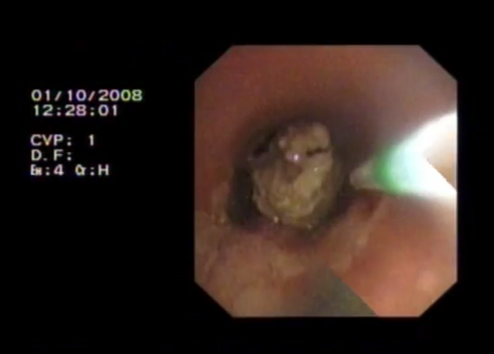
Specialist Bronchoscopic Techniques: the present and the future
Dr Jeremy George treated us to an insight into currently available interventional techniques, of which there are a surprisingly large number, and the possibilities for the near future.
Interventional bronchoscopy is mostly used for large airway obstructive lesions:
- Malignant: primary lung cancer, metastatic lung cancer, thyroid carcinoma, oesophageal carcinoma
- Benign: infection (TB), inflammation (GPA, idiopathic, subglottic stenosis), iatrogenic (post-surgical, intubation injury)
Management is dependant on the underlying cause but for malignant strictures includes:
- tumour debulking: laser resection, diathermy, cryotherapy, blunt dissection with biopsy forceps, and PDT
- intraluminal radiotherapy: indium (decreased rate of regrowth)
- airway stents: silicon stents, self-expanding metal stents (stents are useful for extrinsic compression)
For benign structures options include:
- CO2 laser resection for central strictures
- balloon bronchoplasty +/- depot corticosteroid injections for more peripheral strictures
Laser resection for malignancy is NdYAG, which acts via thermal energy. Beware if you have a plastic ET tube as there is a risk of fire and burnt plastic. To avoid this a rigid bronchoscope can be used, but this requires a GA. A classic paper on the use of laser from the decade of our birth (well, some of us): George, P. J., C. P. Garrett, and M. R. Hetzel. “Role of the neodymium YAG laser in the management of tracheal tumours.” Thorax 42.6 (1987): 440-444.
And a further paper, highlighting that laser can be life saving and allow time to complete surgical procedures: Shankar, S., et al. “Elective resection of tumours of the trachea and main carina after endoscopic laser therapy.” Thorax 45.6 (1990): 493-495.
Dr George described the development of airway stents. They are a foreign body and so are a last resort when other techniques are not possible/unlikely to be effective. In addition, it is important to remember that the complication rates of silicon stents are significant.
- Montgomery, Wiliam W. “T-tube tracheal stent.” Archives of Otolaryngology 82.3 (1965): 320-321.
- Wallace, Michael J., et al. “Tracheobronchial tree: expandable metallic stents used in experimental and clinical applications. Work in progress.” Radiology158.2 (1986): 309-312.
- George, P. J. M., et al. “Covered expandable metal stent for recurrent tracheal obstruction.” The Lancet 335.8689 (1990): 582-584.
- Madden, Brendan P., Subir Datta, and Nick Charokopos. “Experience with Ultraflex expandable metallic stents in the management of endobronchial pathology.” The Annals of thoracic surgery 73.3 (2002): 938-944.
- Wood, Douglas E., et al. “Airway stenting for malignant and benign tracheobronchial stenosis.” The Annals of thoracic surgery 76.1 (2003): 167-174.
Most covered stents cannot be removed, which can be a problem. Late complications of stents include secretion obstruction and stent fracture.
Dr George finished by touching on work at UCLH on surveillance of for the detection of early lung cancer in patients with bronchial dysplasia. Interestingly the natural history of the disease is not simple as not all areas of dysplasia progress to metaplasia and carcinoma. This longitudinal study is allowing us to learn about the natural history of squamous cell carcinoma. Patients with carcinoma in situ or dysplasia but no invasive cancer undergo surveillance with 4-9/12 fluorescence bronchoscopy and annual low dose CT.
Dr George showed videos from his own practice, but if you missed out you can see (inferior) videos on Youtube:
- promotional video on Nd:YAG bronchoscopy
- flexible bronchoscopy airway stent deployment method
- video of multiple bronchoscopic techniques, including airway stent deployment
EBUS-TBNA: why you should be doing it
Dr Bryan Sheinman set out to convince us that all departments of Respiratory Medicine should be doing EBUS-TBNA. He reviewed the history of lung cancer staging techniques, and we remembered a time when patients had to undergo mediastinoscopy for LN sampling. Poor them.
We reviewed the anatomical locations of the relevant LN stations. EBUS-TBNA can reach stations 4R, 4L, 7, 2R, 2L, 10, 11, 12. It cannot reach stations 8 and 9 (for which EUS is required).

We were reminded that at EBUS-TBNA may access the primary mass and LN spread, and therefore identify N2 or N3 disease.
The needle is reassuringly small at 22G. Of course, care must be taken to avoid blood vessels (use of Doppler US aids this), but not too much damage can be done, even if a blood vessel is hit. Reassuring for trainees new to this skill…
EBUS-TBNA results in FNA, not a cell block. However, with modern lab techniques it is possible to characterise and immunostain the ‘worms’ from FNA.
According to NICE EBUS-TBNA has 95% sensitivity and 100% specificity.
Dr Sheinman discussed additional methods of increasing effectiveness, including ROSE. This has significant additional costs and time and staff resources. It is used more often in the private sector. We debated whether the costs were justified. We also debated needs for training, if this technique becomes a standard bronchoscopic skill, rather than an advanced skill as it is currently. A model of fellowships was suggested, but this may not create the requisite number of trained practitioners.
What the physician needs to know about thoracic surgery
Mr Shyam Kolvekar highlighted some key issues in modern approaches to thoracic surgery.
- Open vs VATS: arguments for and against each (see notes on Mr Lau’s talk for the evidence base)
- the various surgical approaches and their advantages and disadvantages
- EBUS has almost entirely replaced mediastinoscopy and future cardiothoracic training may not include mediastinoscopy. It is a blind procedure with a high risk of bleeding and few newly qualified surgeons are keen to get involved
- Pneumonectomy has a 5-10% mortality! A sleeve resection can salvage the lobe and prevent a pneumonectomy being necessary
- For lobectomy FEV1 generally needs to be >1.5L, for pneumonectomy FEV1.2L but these are not concrete. The predicted lung function post-op is what is relevant. TLCO >40% predicted
Modern approaches to Radiotherapy in lung cancer
Dr Julian Singer provided an overview of radiotherapy use in lung cancer.
UK practice for radical RTX in NSCLC:
- stage II and IIIA
- fit patients with PS 0-1 and <10% weight loss
- 55Gy in 20# over 4 weeks (regime chosen for clinical efficacy plus economics)
- 5 year survival 20% (highly selected cases)
USA practice:
- use more fractions (is there a financial incentive to more clinic visits which drives this?)
60Gy vs 74Gy RCT was stopped early as there was too much toxicity in the 74Gy group.
We await the results of an RCT planned for 2015 comparing 60Gy in 30# vs 55Gy in 20# (UK vs USA practice).
Chemotherapy is also given if patient is fit.
Longer treatment tome leads to less toxicity, but more chance of cancer regrowth, therefore treatment given over 4/52 rather than 6/52 gives a better chance of cancer control, but might yield more toxicity to oesophagus, heart, lungs, spinal cord.
Targeted radiotherapy is appropriate for small tumours (T1 or T2A) which are peripheral. This is known as SABR (stereotactic ablative radiotherapy).
- Cyberknife: up to 3cm tumour
- Linac with extra CT built into head to produce 4D CT to plan radiotherapy. So far no RCTs. Single arm trials only but with promising results for peripheral tumours up to 5cm. 1-2% suffer rib #
SABR vs surgery? SABR is given in selected populations and there are no head-to-head trials. Data from the UK SABR consortium gives estimates of local control (1year 92%, 5yr 86%) and survival (1year 82%, 5yr 45%). Trials are needed.
IGRAS: fact and fiction
Prof Onn Min Kon tried to demystify IGRAs and clarify their use and misuse in practice.
Before IGRAs – the TST (Mantoux).
- type IV hypersensitivity reaction
- poor specificity (200 proteins, will respond to BCG and environmental mycobacteria)
- poor sensitivity (10-20% false negative rate in active disease, 25-45% false negative rate in disseminated TB, false negative if CD4 < 200)
- boosting effect of up to 6mm, which leads to problems with repeat testing (eg healthcare workers)
- operator dependant at administration and reading of test
- good historical data
- cheap!
What does MTB have that M. bovis BCG does not? ESAT-6 and CFP-10.
- Renshaw, Philip S., et al. “Structure and function of the complex formed by the tuberculosis virulence factors CFP‐10 and ESAT‐6.” The EMBO journal 24.14 (2005): 2491-2498.
- Behr, M. A., et al. “Comparative genomics of BCG vaccines by whole-genome DNA microarray.” Science 284.5419 (1999): 1520-1523.
- Cole, STea, et al. “Deciphering the biology of Mycobacterium tuberculosis from the complete genome sequence.” Nature 393.6685 (1998): 537-544.
There are currently 2 IGRAs (interferon gamma release assays) available:
- QuantiFERON-TB GOLD: ELISA. £35
- T SPOT TB: enzyme linked immunospot. £50
The former test quantitates the amount of gamma interferon produced in response to the ESAT-6 and CFP-10 antigens from Mycobacterium tuberculosis. The latter test determines the total number of individual effector T cells expressing gamma interferon. IGRAs have high specificity and can be used in BCG-vaccinated individuals to exclude ‘false +ve.’ They have utility as negative predictors of disease in latent TB. NPV high, PPV unclear. They have advantages over the TST, mainly due to the fact that they require only a single visit, and have potential for batching (ideal in outbreak management, new entrant screening, healthcare workers, hard to reach groups).
For latent TB screening there are currently 2 strategies in place (both recommended by NICE): TST then IGRA, or IGRA alone. There is international disagreement on the best strategy in terms of clinical and cost effectiveness.
- Diel, Roland, Robert Loddenkemper, and Albert Nienhaus. “Predictive Value of Interferon-γ Release Assays and Tuberculin Skin Testing for Progression From Latent TB Infection to Disease State A Meta-analysis.” CHEST Journal 142.1 (2012): 63-75.
- Rangaka, Molebogeng X., et al. “Predictive value of interferon-γ release assays for incident active tuberculosis: a systematic review and meta-analysis.” The Lancet infectious diseases 12.1 (2012): 45-55.
- Diel, R., et al. “Interferon-γ release assays for the diagnosis of latent Mycobacterium tuberculosis infection: a systematic review and meta-analysis.”European Respiratory Journal 37.1 (2011): 88-99.
In order to fully assess cost-effectiveness it is not just the initial clinic and visit and the test itself that must be costed. Unnecessary chemoprophylaxis, additional visits and chest x-rays must also be included. TST alone is the most expensive option as it the least specific.
A single step strategy would be optimal if effective.
The risk of progressing to TB if QFN +ve is the same from a smear positive or smear negative index case. However, to complicate the picture, if there is a discordant TST vs IGRA, on re-screening some will have converted, some will have reverted. There is a 50% reversion rate in healthcare workers. Therefore a single step strategy is not the complete answer. The PREDICT study is ongoing, and hopes to provide clarity in this area, assessing the prognostic value of IGRAs in predicting active TB among individuals with, or at risk of, latent TB infection (Dr Marc Lipman at the Royal Free, and Prof Bothamley at Homerton are co-investigators).
What is the place of IGRAs in screening patients with RA planned for TNF-alpha blocking drugs?
Even in those assessed as ‘low risk’ by stratification tables, there were a significant number with positive IGRAs. Stratification plus TST, plus IGRA is recommended to optimise identification of LTBI prior to the use of TNF-alpha antagonists.
Overall, although major progress has been made, when it comes to IGRAs “it’s complicated.”
Treatment of OSA
Dr Himender Makker covered new aspects of sleep apnoea.
OSA occurs because the upper airway is narrow and collapsible.
CPAP is the gold standard treatment, and was developed by Dr Colin Sullivan who published his n= 5 study in the Lancet in 1981. A Cochrane review of 2006 and guidance from NICE in 2008 recommend the treatment.
- Sullivan, ColinE, et al. “Reversal of obstructive sleep apnoea by continuous positive airway pressure applied through the nares.” The Lancet 317.8225 (1981): 862-865.
- Giles, Tammie L., et al. “Continuous positive airways pressure for obstructive sleep apnoea in adults.” Cochrane Database Syst Rev 3.3 (2006).
- McDaid, Catriona, et al. “Continuous positive airway pressure devices for the treatment of obstructive sleep apnoea–hypopnoea syndrome: a systematic review and economic analysis.” (2009).
ENT surgery including tonsillectomy, and UPPP has varioable outcomes but is not reliable as the obstruction is not at a single level. It should only be recommended for highly selected patients where the obstruction can be shown to be localised. We were reminded that tracheostomy is a highly effective treatment for OSA, if a little drastic!
Other treatment modalities such as upper airway remodelling by orofacial exercise have also been proposed, with mixed results so far.
Not sure what orofacial exercises involve? Youtube has the answer.
How to write a business case
We finished the day with advice from Penny O’Hara on how to write a business case, an essential skill for any Consultant. She provided some top tips:
- A business case is a collaborative effort, so find allies amongst clinical colleagues and management
- Try to understand different perspectives: financial, informatics, management
- Marketing is key. You need to not only justify, but sell your idea
- Align with the Trust strategy and you’re much more likely to be successful
- Practice presenting your argument as you need to be coherent and persuasive
- Personal relationships are important; acknowledge and cultivate them
- Keep your message simple
Her presentation is available here: How to write a business case April 2014V7Final
See you all soon.

Discussion
No comments yet.