In September we gathered at Basildon Hospital for a day of CPET, Radiology and pleural disease.
CPET in breathlessness
Dr Johnson Samuel started the day with a case of breathlessness in which standard tests were normal. A 40 year old man was short of breath on cycling. He was found to have a normal chest x-ray and spirometry. A CPET showed that his heart rate did not rise with exercise and he was diagnosed with primary chronotropic incompetence (autonomic dysfunction). Dr Johnson highlighted that resting tests cannot reliably predict exercise performance.
The differential diagnosis for unexplained breathlessness is wide: dysfunctional breathing, arrhythmia, neuromuscular weakness, obesity, pulmonary vascular disease, exercise-induced asthma, LV diastolic dysfunction, early myocardial ischaemia, R to L shunt, and deconditioning. CPET can assess the response to exercise in an integrated way, and identify the anaerobic threshold. Measurements are taken of: VO2, VCO2, HR, ECG, Sats, Blood gas. Plotting data allows interpretation which aids identification of cardiovascular limitation vs respiratory limitation vs VQ mismatch vs peripheral system problems.
A further illustrative case detailed a 35 year old PE teacher with dysfunctional breathing. On the CPET transient T wave changes were seen on ECG due to hypoxia/psychogenic dyspnoea. Exclusion of other causes of breathlessness aided management and allowed the man to return to work reassured.
Radiology for SpRs
Dr A Anilkumar took us through PET-CT, highlighting its value as both an anatomical and functional investigation. We were reminded that PET is based on positron e+ with FDG as the marker. We were also reminded of the radiation dose of PET-CT as compared to other investigations and background radiation:
- PET-CT = 18msv
- CT Chest = 6.6msv
- CXR = 0.02msv
- Annual radiation dose in Cornwall = 7.8msv
- Annual radiation dose in rest of UK = 2.7msv
Interpretation of PET/CT requires expertise. There are a number of normal variants that can cause ‘hot spots’ including brown fat, muscle uptake, LV, brain, liver, kidneys and bladder, larynx and stomach. The colon is also a difficult area of interpretation on PET.
RCR indications for PET-CT include:
- lung cancer staging if considered for radical treatments (NSCLC or limited SCLC)
- SPN 10mm+
- failed biopsy/technically difficult biopsy
- suspected disease recurrence after lung cancer treatment
- pleural malignancy (to guide biopsy and exclude extrathoracic disease)
Positive on PET means SUV>2.5. False positives include: TB, rheumatoid nodule, fungal infection, granulomata. False negatives include: pulmonary carcinoid, BAC, < 6mm and micrometastases. PET has good sensitivity for adrenal mets, but poor for brain mets. Contrast enhanced PET-CT is helpful for invasion ie T stage.
A positive node on PET needs EBUS. If this is negative it can down-stage the cancer which may change treatment options.
Pleural effusion management
Dr Marcus Pittman encouraged us all to read the BTS guidelines on pleural effusion investigation and management, and ran through the algorithm.
Ambulatory pathways and PleurX
Dr Kirsten Wadsworth introduced ambulatory care pathways for patients with pleural effusions. Where these are established they are predominantly for malignant effusions. The aim is to keep patients out f hospital, save bed days, decreased costs and increase patient satisfaction.
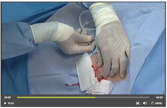
This handy video shows how to insert a PleurX catheter. It features the obligatory patient walking on the beach, miraculously free of symptoms…
Currently indwelling drains are manly used for failed pleurodesis or continuous drainage. Some advocate for indwelling catheters as first line treatment for malignant effusions. Patient preference has not been established.
Talc Pleurodesis
Dr Kanwar Pannu examined myths and realities of talc pleurodesis. He reminded us that historically talc was 5-10microns and was associated with a risk of complications such as respiratory failure. However talc of higher particle size (ie 31.3 microns French mean) is associated with fewer complications. Talc slurry leads to an intense inflammatory reaction, and therefore steroids should be crossed off if possible, as they counteract this inflammation. It is advisable to stop NSAIDS for 48 hour pre and post-procedure. There is evidence that stopping diclofenac leads to better outcomes, and this is likely to be a class effect.
Talc slurry should be attempted when drainage is <150ml/24hr or 90% resolution on CXR. A 2006 meta-analysis showed no evidence for tilting/rolling patients post-talc slurry. In addition there was no difference in outcome at 30 days for talc poudrage vs talc slurry. VATS is not recommended for malignant effusion as there is no significant difference in outcome and potential for more complications.
This is different for pneumothorax, in which an initial safety trial showed a favourable outcome for thoracoscopic talc pleurodesis in recurrent primary spontaneous pneumothorax.
Medical Thoracoscopy
Dr Dipak Mukherjee pitched Basildon Hospital as a great place for trainees and Consultants to work. He described the roles of the 6 Consultants, 4 middle grades and 2 SpRs. He highlighted the services provided at Basildon, including home NIV, CPET, EBUS, and Medical Thoracoscopy (cryotherapy and endobronchial diathermy coming soon).
Medical thoracoscopy requires skills in conscious sedation. Basildon favours midazolam and pethidine. Alternatives are fentanyl and alfentanyl. Propofol is also an option but is used less. The patient in placed in the lateral decubitus position and access is gained via the safe triangle via skin incision and blunt dissection. Interestingly diseased pleura is less sensitive/painful than normal pleura so medical thoracoscopy is well-tolerated. Local anaesthesia is used +/- pethidine and post-procedure analgesia. Some favour 1 port, other 2 ports. Mortality is no higher than 0.24% which is similar to transbronchial biopsy.
As with all procedures, patient selection is key to good outcomes.
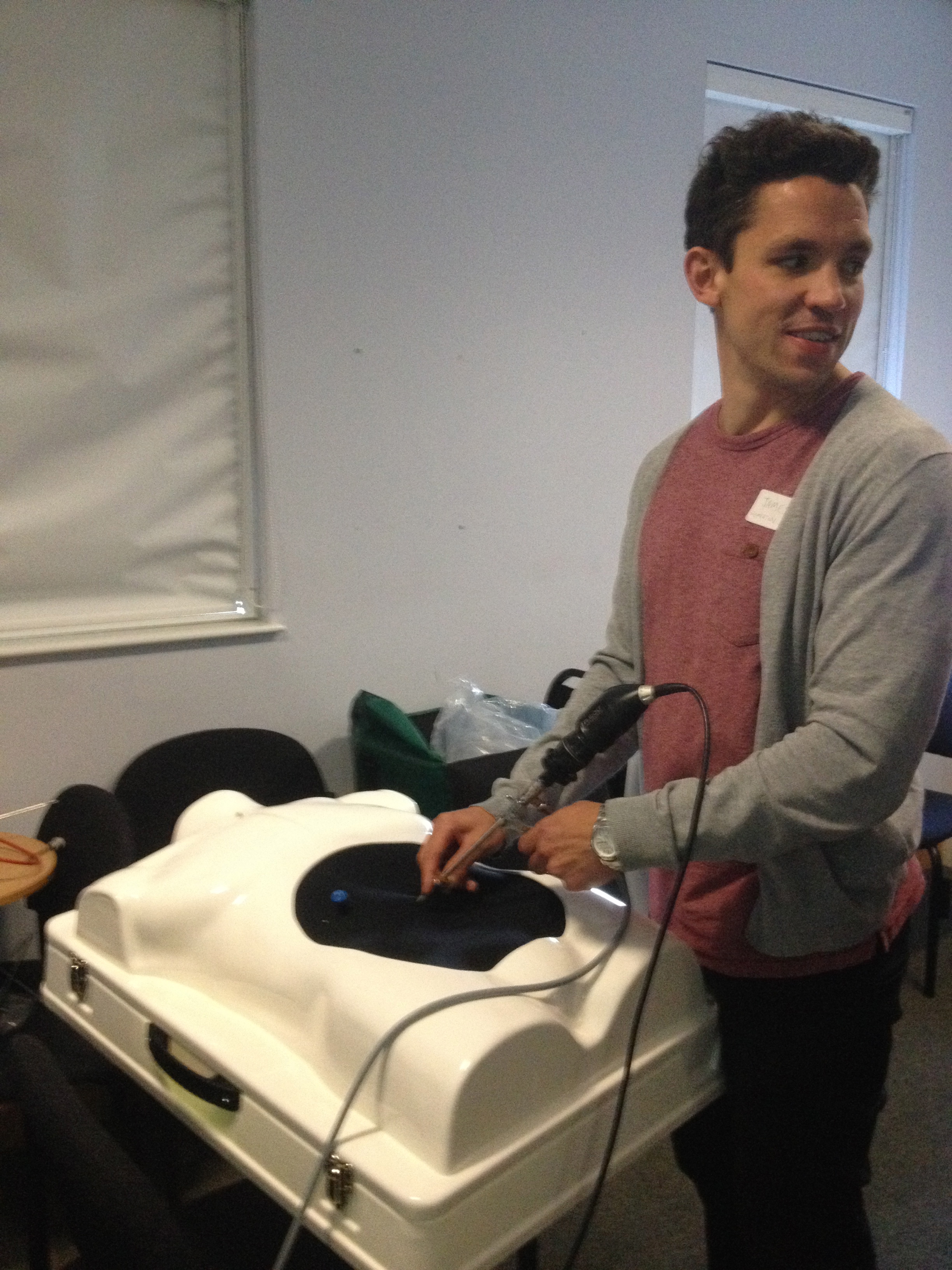

In the afternoon there was an opportunity for some hand-on experience of a thoracoscope, using a training model.
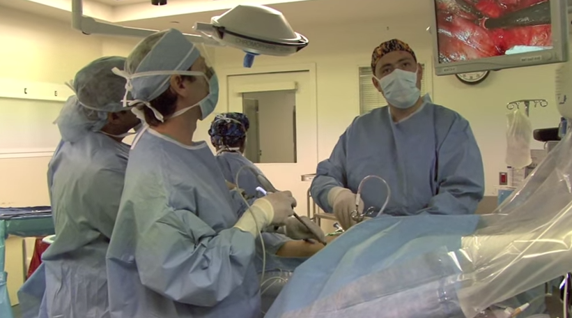
And there was also a valiant but ultimately unsuccessful attempt to use video link to theatres to watch a VATS. If you missed this, why not watch one on YouTube?
We hope to see you at the next training day.

Discussion
No comments yet.